Clinical Pathways for the 5 Wound Types
Choose a wound type to view the associated clinical pathways and guidelines.
Arterial Ulcer Pathway
Arterial Ulcer:
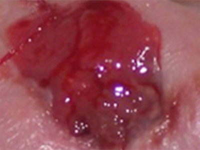
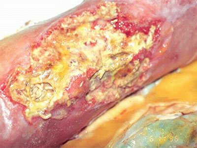
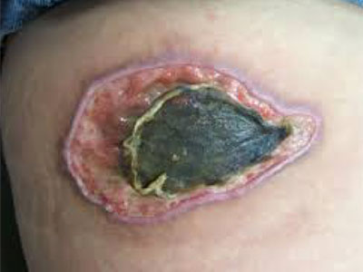
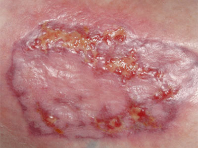
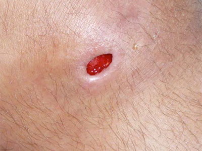
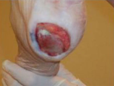
 Patient with ulcer located distally on the lower extremity, most often seen on the forefoot or toes. Typically with dry, punched out appearance, usually on a non weight bearing surface.
Patient with ulcer located distally on the lower extremity, most often seen on the forefoot or toes. Typically with dry, punched out appearance, usually on a non weight bearing surface.
Step 1
Assess:
- Presence and quality of pulse
- Color, temperature
- Toe nail thickening
- Hair loss
- Thin flaky skin
- Shiny skin
Step 2
Obtain:
- Vascular history
- Surgical history
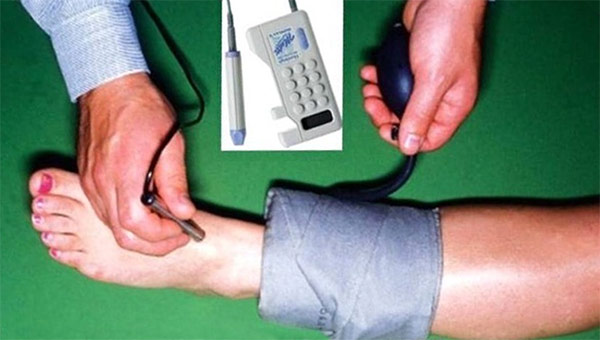
- ABI
Step 3
Nutritional Status:
- Order “wound panel”
- Optimize blood glucose level
- Assess other host factors
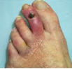
Signs/Symptoms of Infection Present?
No:
- Paint with betadine or skin barrier daily
- Apply dry gauze or clean cotton socks daily
- Monitor closely for signs/symptoms of infection
Signs/Symptoms of Infection Present?
Yes, with nonviable tissue:
- Consider systemic antibiotics
- Refer to wound clinic for debridement
- Choose debridement product while waiting for clinic appointment
Signs/Symptoms of Infection Present?
Yes, with viable tissue:
- Consider systemic antibiotics
- Consider silver product
References
Ruth A. Bryant, Denise P. Nix (2012), Acute and chronic wounds - Current management concepts (4th edition). St. Louis, MO: Elsevier Mosby
Emory University WOC Education Program (2012), Skin and Wound Module. Atlanta, GA: Emory University WOCNEC